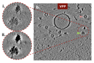
The Volta phase plate (VPP) can enhance low-frequency phase contrast in cryo-electron microscopy (cryo-EM) and cryo-electron tomography (cryo-ET), but its impact on tomogram alignment has not been systematically assessed. Using sub-2Å single-particle analysis (SPA) and analytical contrast transfer function (CTF) modeling, we show that despite high-frequency information loss and moderate signal damping, the VPP enhances signal in low-frequency bands that are crucial for tilt-series alignment. We show that VPP tomograms exhibit increased contrast and remain higher in contrast post denoising, for thick and crowded specimens. Quantitative comparison of tomograms acquired with and without the VPP shows improved tilt-axis orientation measurements and a higher fraction of tomograms amenable to local tilt-series alignment. Although acquisition with the VPP yields lower subtomogram averaging (STA) resolution, these results show that the primary benefits of the VPP for cryo-ET are contrast enhancement and improved tilt-series alignment robustness, which are crucial for studying cellular ultrastructure and protein organization in crowded environments, where STA at moderate resolutions remains valuable.
@article{hutchings2026volta,
  author = {Hutchings, Joshua and Ji, Daniel and Zheng, Shawn and Montabana, Elizabeth A. and Ermel, Utz H. and Peck, Ariana and Schwartz, Jonathan and Woldeyes, Rahel and Paraan, Mohammadreza and Ali, Mallak and Hill, Norbert S. and Siems, Hannah and Serwas, Daniel and Cheng, Anchi and Kimanius, Dari and Agard, David A. and Potter, Clinton S. and Carragher, Bridget and Yu, Yue},
  title = {Evaluating the Volta phase plate for improved tomogram alignment in cryo-electron tomography},
  journal = {IUCrJ},
  year = {2026},
  volume = {13},
  number = {3},
  month = {May},
  doi = {10.1107/S2052252526002575},
}
2025
Cryo-ET
A Realistic Phantom Dataset for Benchmarking Cryo-ET Data Annotation
Ariana Peck, Yue Yu, Jonathan Schwartz, and
17 more authors
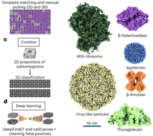
Cryo-electron tomography (cryo-ET) is a powerful technique for imaging molecular complexes in their native cellular environments. However, identifying the vast majority of molecular species in cellular tomograms remains prohibitively difficult. Machine learning (ML) methods provide an opportunity to automate the annotation process, but algorithm development has been hindered by the lack of large, standardized datasets. Here we present an experimental phantom dataset with comprehensive ground-truth annotations for six molecular species to spur new algorithm development and benchmark existing tools. This annotated dataset is available on the CryoET Data Portal with infrastructure to streamline access for methods developers across fields.
@article{peck2025phantom,
  title = {A Realistic Phantom Dataset for Benchmarking Cryo-ET Data Annotation},
  author = {Peck, Ariana and Yu, Yue and Schwartz, Jonathan and Cheng, Anchi and Ermel, Utz Heinrich and Hutchings, Joshua and Kandel, Saugat and Kimanius, Dari and Montabana, Elizabeth A. and Serwas, Daniel and Siems, Hannah and Wang, Feng and Zhao, Zhuowen and Zheng, Shawn and Haury, Matthias and Agard, David A. and Potter, Clinton S. and Carragher, Bridget and Harrington, Kyle and Paraan, Mohammadreza},
  journal = {Nature Methods},
  volume = {22},
  number = {9},
  pages = {1819--1823},
  year = {2025},
  doi = {10.1038/s41592-025-02800-5},
}
Machine Learning
Optimal 3D Chemical Imaging with Multimodal Electron Tomography
Jason Manassa, William Millsaps, Jonathan Schwartz, and Robert Hovden
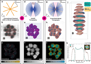
Accurate mapping of nanoscale chemistry in three dimensions (3D) has been a longstanding challenge. Modern electron microscopy provides chemical images by electron energy loss spectroscopy (EELS) and energy dispersive x-ray spectrometry (EDX) but requires high fluences that damage specimens. In 3D, the requirements are worse; electron tomography demands many high-fluence chemical maps for reconstruction, creating a tradeoff between resolution, accuracy, and sample survival. Fused multimodal electron tomography (MM-ET) alleviates this requirement by leveraging lower-fluence high-angle annular dark-field (HAADF) images alongside a few chemical maps to dramatically improve chemical resolution. Here, experimental and computational parameter space is systematically explored to determine when MM-ET performs best. Ideal imaging conditions balance sample survival with resolution and chemical specificity; we recommend a tilt range of at least ± 70∘, acquiring 40 equally spaced HAADF projections (signal-to-noise > 10), and 7 EELS/EDX maps of each chemistry (signal-to-noise > 4).
@article{manassa2025optimal,
  title = {Optimal 3D Chemical Imaging with Multimodal Electron Tomography},
  author = {Manassa, Jason and Millsaps, William and Schwartz, Jonathan and Hovden, Robert},
  journal = {npj Computational Materials},
  volume = {11},
  number = {1},
  year = {2025},
  doi = {10.1038/s41524-025-01750-y},
}
2024
Inverse Problems
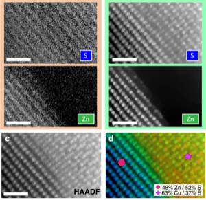
Imaging 3D Chemistry at 1 nm Resolution with Fused Multi-Modal Electron Tomography
Jonathan Schwartz, Zichao Wendy Di, Yi Jiang, and
14 more authors
Measuring the three-dimensional (3D) distribution of chemistry in nanoscale matter is a longstanding challenge for metrological science. The inelastic scattering events required for 3D chemical imaging are too rare, requiring high beam exposure that destroys the specimen before an experiment is completed. Even larger doses are required to achieve high resolution. Thus, chemical mapping in 3D has been unachievable except at lower resolution with the most radiation-hard materials. Here, high-resolution 3D chemical imaging is achieved near or below one-nanometer resolution in an Au-Fe3O4 metamaterial within an organic ligand matrix, Co3O4-Mn3O4 core-shell nanocrystals, and ZnS-Cu0.64S0.36 nanomaterial using fused multi-modal electron tomography. Multi-modal data fusion enables high-resolution chemical tomography often with 99% less dose by linking information encoded within both elastic (HAADF) and inelastic (EDX/EELS) signals. We thus demonstrate that sub-nanometer 3D resolution of chemistry is measurable for a broad class of geometrically and compositionally complex materials.
@article{yang2024imaging3d,
  title = {Imaging 3D Chemistry at 1 nm Resolution with Fused Multi-Modal Electron Tomography},
  author = {Schwartz, Jonathan and Di, Zichao Wendy and Jiang, Yi and Manassa, Jason and Pietryga, Jacob and Qian, Yiwen and Cho, Min Gee and Rowell, Jonathan L. and Zheng, Huihuo and Robinson, Richard D. and Gu, Junsi and Kirilin, Alexey and Rozeveld, Steve and Ercius, Peter and Fessler, Jeffrey A. and Scott, Mary and Hovden, Robert},
  journal = {Nature Communications},
  volume = {15},
  pages = {47558},
  year = {2024},
  doi = {10.1038/s41467-024-47558-0},
}
Materials
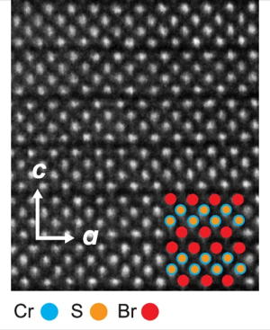
Extraordinary Phase Transition Revealed in a van der Waals Antiferromagnet
Xiaoyu Guo, Wenhao Liu, Jonathan Schwartz, and
12 more authors
While the surface-bulk correspondence has been ubiquitously shown in topological phases, the relationship between surface and bulk in Landau-like phases is much less explored. Theoretical investigations since 1970s for semi-infinite systems have predicted the possibility of the surface order emerging at a higher temperature than the bulk, clearly illustrating a counterintuitive situation and greatly enriching phase transitions. But experimental realizations of this prediction remain missing. Here, we demonstrate the higher-temperature surface and lower-temperature bulk phase transitions in CrSBr, a van der Waals (vdW) layered antiferromagnet. We leverage the surface sensitivity of electric dipole second harmonic generation (SHG) to resolve surface magnetism, the bulk nature of electric quadrupole SHG to probe bulk spin correlations, and their interference to capture the two magnetic domain states. Our density functional theory calculations show the suppression of ferromagnetic-antiferromagnetic competition at the surface is responsible for this enhanced surface magnetism. Our results not only show counterintuitive, richer phase transitions in vdW magnets, but also provide viable ways to enhance magnetism in their 2D form.
@article{guo2024extraordinary,
  title = {Extraordinary Phase Transition Revealed in a van der Waals Antiferromagnet},
  author = {Guo, Xiaoyu and Liu, Wenhao and Schwartz, Jonathan and Sung, Suk Hyun and Zhang, Dechen and Shimizu, Makoto and Kondusamy, Aswin L. N. and Li, Lu and Sun, Kai and Deng, Hui and Jeschke, Harald O. and Mazin, Igor I. and Hovden, Robert and Lv, Bing and Zhao, Liuyan},
  journal = {Nature Communications},
  volume = {15},
  pages = {6472},
  year = {2024},
  doi = {10.1038/s41467-024-50900-1},
}
2023
Machine Learning
Autonomous Electron Tomography Reconstruction with Machine Learning
William Millsaps, Jonathan Schwartz, Zichao Wendy Di, Yi Jiang, and Robert Hovden
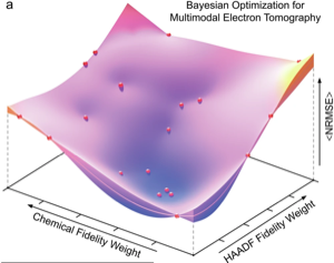
Modern electron tomography has progressed to higher resolution at lower doses by leveraging compressed sensing (CS) methods that minimize total variation (TV). However, these sparsity-emphasized reconstruction algorithms introduce tunable parameters that greatly influence the reconstruction quality. Here, Pareto front analysis shows that high-quality tomograms are reproducibly achieved when TV minimization is heavily weighted. However, in excess, CS tomography creates overly smoothed three-dimensional (3D) reconstructions. Adding momentum to the gradient descent during reconstruction reduces the risk of over-smoothing and better ensures that CS is well behaved. For simulated data, the tedious process of tomography parameter selection is efficiently solved using Bayesian optimization with Gaussian processes. In combination, Bayesian optimization with momentum-based CS greatly reduces the required compute time—an 80% reduction was observed for the 3D reconstruction of SrTiO nanocubes. Automated parameter selection is necessary for large-scale tomographic simulations that enable the 3D characterization of a wider range of inorganic and biological materials.
@article{millsaps2023autonomous,
  title = {Autonomous Electron Tomography Reconstruction with Machine Learning},
  author = {Millsaps, William and Schwartz, Jonathan and Di, Zichao Wendy and Jiang, Yi and Hovden, Robert},
  journal = {Microscopy and Microanalysis},
  volume = {29},
  number = {5},
  pages = {1650--1659},
  year = {2023},
}
Materials
Photonically Active Bowtie Nanoassemblies with Chirality Continuum
N. Kotov, P. Kumar, T. Vo, M. Cha, A. Visheratina, J.K. Kim, W. Xu, Jonathan Schwartz, et al.
Chirality is a geometrical property described by continuous mathematical functions. However, in chemical disciplines, chirality is often treated as a binary left or right characteristic of molecules rather than a continuity of chiral shapes. Although they are theoretically possible, a family of stable chemical structures with similar shapes and progressively tuneable chirality is yet unknown. Here we show that nanostructured microparticles with an anisotropic bowtie shape display chirality continuum and can be made with widely tuneable twist angle, pitch, width, thickness and length. The self-limited assembly of the bowties enables high synthetic reproducibility, size monodispersity and computational predictability of their geometries for different assembly conditions. The bowtie nanoassemblies show several strong circular dichroism peaks originating from absorptive and scattering phenomena. Unlike classical chiral molecules, these particles show a continuum of chirality measures that correlate exponentially with the spectral positions of the circular dichroism peaks. Bowtie particles with variable polarization rotation were used to print photonically active metasurfaces with spectrally tuneable positive or negative polarization signatures for light detection and ranging (LIDAR) devices.
@article{kotov2023photonically,
  title = {Photonically Active Bowtie Nanoassemblies with Chirality Continuum},
  author = {Kotov, Nicholas and Kumar, Prashant and Vo, Thi and Cha, Minjeong and Visheratina, Anastasiia and Kim, J.K. and Xu, Wei and Schwartz, Jonathan and others},
  journal = {Nature},
  volume = {615},
  pages = {418--424},
  year = {2023},
}
2022
Materials
GaN-based Deep-nano Structures: Break the Efficiency Bottleneck of Conventional Nanoscale Optoelectronics
A. Navid, A. Pandey, Y.M. Goh, Jonathan Schwartz, R. Hovden, Z. Mi
Conventional semiconducting nanowire optoelectronic devices generally exhibit low efficiency, due to dominant nonradiative surface recombination. Here, it is shown that such a critical challenge can be potentially addressed by exploiting semiconducting structures in the deep-nanoregime. The epitaxy and structural and optical characteristics of GaN-based micro-network nanostructures grown on Si wafer are studied. These complex nanostructures have lateral dimensions as small as a few nanometers. Detailed scanning transmission electron microscopy studies suggest that the self-assembled micro-network nanostructures are monocrystalline, despite the porous nature. Significantly, such micro-network nanostructures exhibit ultrabright emission in the visible spectrum. Compared to conventional InGaN nanowire structures with similar surface area, the surface recombination velocity of such deep-nanostructures is reduced by nearly two orders of magnitude, which is evidenced by the extremely bright luminescence emission as well as the long carrier lifetime measured under low excitation conditions. This study offers a new path for the design and development of next generation high efficiency nanoscale optoelectronic devices.
@article{navid2022gan,
  title = {GaN-based Deep-nano Structures: Break the Efficiency Bottleneck of Conventional Nanoscale Optoelectronics},
  author = {Navid, Alireza and Pandey, Ayush and Goh, Yeow Ming and Schwartz, Jonathan and Hovden, Robert and Mi, Zetian},
  journal = {Advanced Optical Materials},
  pages = {2102263},
  year = {2022},
}
Sci. Software
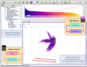
Real-time 3D Analysis During Electron Tomography Using tomviz
Jonathan Schwartz, Chris Harris, Jacob Pietryga, and
14 more authors
The demand for high-throughput electron tomography is rapidly increasing in biological and material sciences. However, this 3D imaging technique is computationally bottlenecked by alignment and reconstruction which runs from hours to days. We demonstrate real-time tomography with dynamic 3D tomographic visualization to enable rapid interpretation of specimen structure immediately as data is collected on an electron microscope. Using geometrically complex chiral nanoparticles, we show volumetric interpretation can begin in less than 10 minutes and a high-quality tomogram is available within 30 minutes. Real-time tomography is integrated into tomviz, an open-source and cross-platform 3D data analysis tool that contains intuitive graphical user interfaces (GUI), to enable any scientist to characterize biological and material structure in 3D.
@article{schwartz2022real,
  title = {Real-time 3D Analysis During Electron Tomography Using tomviz},
  author = {Schwartz, Jonathan and Harris, Chris and Pietryga, Jacob and Zheng, Huihuo and Kumar, Prashant and Visheratina, Anastasiia and Kotov, Nicholas A. and Major, Brianna and Avery, Patrick and Ercius, Peter and Ayachit, Utkarsh and Geveci, Berk and Muller, David A. and Genova, Alessandro and Jiang, Yi and Hanwell, Marcus and Hovden, Robert},
  journal = {Nature Communications},
  volume = {13},
  number = {1},
  pages = {4458},
  year = {2022},
  doi = {10.1038/s41467-022-32046-0},
}
Inverse Problems
Imaging Atomic-Scale Chemistry from Fused Multi-Modal Electron Microscopy
Jonathan Schwartz, Zichao Wendy Di, Yi Jiang, and
9 more authors
Efforts to map atomic-scale chemistry at low doses with minimal noise using electron microscopes are fundamentally limited by inelastic interactions. Here, fused multi-modal electron microscopy offers high signal-to-noise ratio (SNR) recovery of material chemistry at nano- and atomic-resolution by coupling correlated information encoded within both elastic scattering (high-angle annular dark-field (HAADF)) and inelastic spectroscopic signals (electron energy loss (EELS) or energy-dispersive x-ray (EDX)). By linking these simultaneously acquired signals, or modalities, the chemical distribution within nanomaterials can be imaged at significantly lower doses with existing detector hardware. In many cases, the dose requirements can be reduced by over one order of magnitude. This high SNR recovery of chemistry is tested against simulated and experimental atomic resolution data of heterogeneous nanomaterials.
@article{mi2022imaging,
  title = {Imaging Atomic-Scale Chemistry from Fused Multi-Modal Electron Microscopy},
  author = {Schwartz, Jonathan and Di, Zichao Wendy and Jiang, Yi and Fielitz, Alyssa J. and Ha, Don-Hyung and Perera, Sanjaya D. and El Baggari, Ismail and Robinson, Richard D. and Fessler, Jeffrey A. and Ophus, Colin and Rozeveld, Steve and Hovden, Robert},
  journal = {npj Computational Materials},
  volume = {8},
  pages = {16},
  year = {2022},
  doi = {10.1038/s41524-021-00692-5},
}
Materials
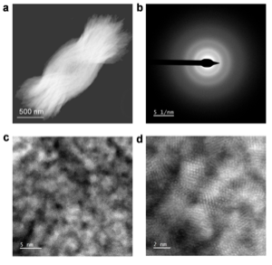
A Three-Stage Magnetic Phase Transition Revealed in Ultrahigh-Quality van der Waals Bulk Magnet CrSBr
Wenhao Liu, Xiaoyu Guo, Jonathan Schwartz, and
10 more authors
van der Waals (vdW) magnets are receiving ever-growing attention nowadays due to their significance in both fundamental research on low-dimensional magnetism and potential applications in spintronic devices. The high crystalline quality of vdW magnets is the key to maintaining intrinsic magnetic and electronic properties, especially when exfoliated down to the two-dimensional limit. Here, ultrahigh-quality air-stable vdW CrSBr crystals are synthesized using the direct solid–vapor synthesis method. The high single crystallinity and spatial homogeneity have been thoroughly evidenced at length scales from submm to atomic resolution by X-ray diffraction, second harmonic generation, and scanning transmission electron microscopy. More importantly, specific heat measurements of ultrahigh-quality CrSBr crystals show three thermodynamic anomalies at 185, 156, and 132 K, revealing a stage-by-stage development of the magnetic order upon cooling, which is also corroborated with the magnetization and transport results. Our ultrahigh-quality CrSBr can further be exfoliated down to monolayers and bilayers easily, providing the building blocks of heterostructures for spintronic and magneto-optoelectronic applications.
@article{liu2022three,
  title = {A Three-Stage Magnetic Phase Transition Revealed in Ultrahigh-Quality van der Waals Bulk Magnet CrSBr},
  author = {Liu, Wenhao and Guo, Xiaoyu and Schwartz, Jonathan and Xie, Hongchao and Dhale, Nikhil Uday and Sung, Suk Hyun and Kondusamy, Aswin Lakshmi Narayanan and Wang, Xiqu and Zhao, Haonan and Berman, Diana and Hovden, Robert and Zhao, Liuyan and Lv, Bing},
  journal = {ACS Nano},
  volume = {16},
  number = {10},
  pages = {15917--15926},
  year = {2022},
  doi = {10.1021/acsnano.2c02896},
}
Materials
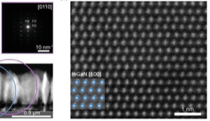
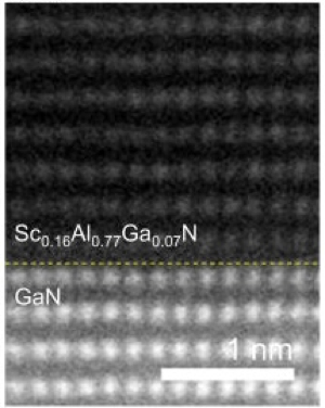
Quaternary Alloy ScAlGaN: A Promising Strategy to Improve the Quality of ScAlN
Ping Wang, Ding Wang, Yutong Bi, Boyu Wang, Jonathan Schwartz, Robert Hovden, and Zetian Mi
ScAlN is an emerging ultrawide bandgap semiconductor for next-generation radio frequency electronic devices. Here, we show that the material quality of ScAlN grown by molecular beam epitaxy can be drastically improved by alloying with Ga. The resulting quaternary alloy ScAlGaN exhibits a single-phase wurtzite structure, atomically smooth surface, high crystal quality, sharp interface, and low impurity concentration. Most significantly, oxygen impurity incorporation in ScAlGaN is found to be three to four orders of magnitude lower compared to that for ScAlN grown on AlN templates utilizing a similar Sc source. We further demonstrate that ScAlGaN/GaN superlattices exhibit clear periodicity with sharp interfaces. Moreover, GaN high electron mobility transistors with high sheet electron density and high mobility have been realized using ScAlGaN as a barrier. This work provides a viable approach for achieving high-quality Sc-III-N semiconductors that were not previously possible and further offers additional dimensions for bandgap, polarization, interface, strain, and quantum engineering.
@article{wang2022scalgan,
  title = {Quaternary Alloy ScAlGaN: A Promising Strategy to Improve the Quality of ScAlN},
  author = {Wang, Ping and Wang, Ding and Bi, Yutong and Wang, Boyu and Schwartz, Jonathan and Hovden, Robert and Mi, Zetian},
  journal = {Applied Physics Letters},
  volume = {120},
  number = {1},
  pages = {012104},
  year = {2022},
  doi = {10.1063/5.0060608},
}
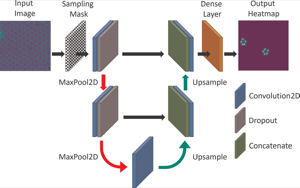
Deep Learning
Atomic Defect Identification with Sparse Sampling and Deep Learning
Michael C. Cao, Jonathan Schwartz, Huihuo Zheng, Yi Jiang, Robert Hovden, and Yimo Han
Communications in Computer and Information Science, 2022
Scanning Transmission Electron Microscopy (STEM) is a high-resolution characterization technique that can resolve atomic lattices. Recent advances in high-brightness sources enable in-situ STEM experiments with acquisition rates reaching 20 frames per second. However, high-doses of electron radiation damage the atomic lattice and limit frame-rates. Thus the real-time visualization of lattice transformations with sub-angstrom resolution requires innovative tools to track defect evolution while limiting electron radiolysis. Here we present a trained deep learning model that automatically tracks lattice defects in graphene while scanning around 60% of the total area for an in-situ experiment. Our approach extracts relevant physical information from STEM movies without requiring any knowledge of the lattice symmetry. Atomic defect identification using sparse sampling and deep learning was demonstrated on a multi-image dataset of defects in graphene as part of the 2021 Smoky Mountains Data Challenge.
@inproceedings{cao2022atomic,
  title = {Atomic Defect Identification with Sparse Sampling and Deep Learning},
  author = {Cao, Michael C. and Schwartz, Jonathan and Zheng, Huihuo and Jiang, Yi and Hovden, Robert and Han, Yimo},
  booktitle = {Communications in Computer and Information Science},
  volume = {1512},
  pages = {455--463},
  year = {2022},
  doi = {10.1007/978-3-030-96498-6_28},
  publisher = {Springer},
}
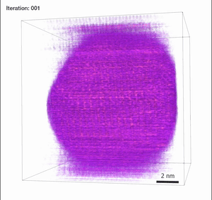
2020
Inverse Problems
Dynamic Compressed Sensing for Real-Time Tomographic Reconstruction
Huihuo Zheng, Jonathan Schwartz, Marcus Hanwell, Yi Jiang, and Robert Hovden
Electron tomography has achieved higher resolution and quality at reduced doses with recent advances in compressed sensing. Compressed sensing (CS) exploits the inherent sparse signal structure to efficiently reconstruct three-dimensional (3D) volumes at the nanoscale from undersampled measurements. However, the process bottlenecks 3D reconstruction with computation times that run from hours to days. Here we demonstrate a framework for dynamic compressed sensing that produces a 3D specimen structure that updates in real-time as new specimen projections are collected. Researchers can begin interpreting 3D specimens as data is collected to facilitate high-throughput and interactive analysis. Using scanning transmission electron microscopy (STEM), we show that dynamic compressed sensing accelerates the convergence speed by ~3-fold while also reducing its error by 27% for a Au/SrTiO3 nanoparticle specimen. Before a tomography experiment is completed, the 3D tomogram has interpretable structure within ~33% of completion and fine details are visible as early as ~66%. Upon completion of an experiment, a high-fidelity 3D visualization is produced without further delay. Additionally, reconstruction parameters that tune data fidelity can be manipulated throughout the computation without re-running the entire process.
@article{zheng2020dynamic,
  title = {Dynamic Compressed Sensing for Real-Time Tomographic Reconstruction},
  author = {Zheng, Huihuo and Schwartz, Jonathan and Hanwell, Marcus and Jiang, Yi and Hovden, Robert},
  journal = {Ultramicroscopy},
  volume = {219},
  pages = {113122},
  year = {2020},
  doi = {10.1016/j.ultramic.2020.113122},
}
2019
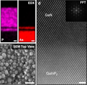
Materials
Stable Unassisted Solar Water Splitting on Semiconductor Photocathodes Protected by Multifunctional GaN Nanostructures
Yongjie Wang, Jonathan Schwartz, Jiseok Gim, Robert Hovden, and Zetian Mi
Producing hydrogen by unassisted solar water splitting is one essential step to make direct solar fuel conversion a viable energy source. To date, however, there has been no demonstration of stable photoelectrodes for high-efficiency photoelectrochemical water splitting. In this work, we report that a GaInP2/GaAs/Ge triple-junction (3J) photocathode protected by multifunctional GaN nanostructures can enable both efficient and relatively stable solar water splitting. A 12.6% solar-to-hydrogen (STH) efficiency is measured without any external bias. Of particular importance, we demonstrate relatively stable solar water splitting for 80 h in three-electrode configuration and 57 h in two-electrode measurement at zero bias. This is the best reported stability for multijunction III-V semiconductor photocathodes in two-electrode configuration to our knowledge. The multifunctional GaN nanostructure significantly reduces the charge transfer resistance at the semiconductor/electrolyte interface and protects III-V materials against corrosion. Such multifunctional GaN photocatalytic nanostructures provide a new pathway to improve the performance of conventional photoelectrodes to achieve both efficient and stable unassisted solar water splitting.
@article{wang2019stable,
  title = {Stable Unassisted Solar Water Splitting on Semiconductor Photocathodes Protected by Multifunctional GaN Nanostructures},
  author = {Wang, Yongjie and Schwartz, Jonathan and Gim, Jiseok and Hovden, Robert and Mi, Zetian},
  journal = {ACS Energy Letters},
  volume = {4},
  number = {7},
  pages = {1541--1548},
  year = {2019},
  doi = {10.1021/acsenergylett.9b00549},
}
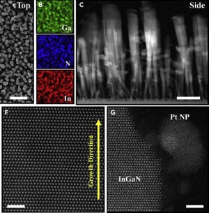
Materials
A Single-Junction Cathodic Approach for Stable Unassisted Solar Water Splitting
Yongjie Wang, Yuanpeng Wu, Jonathan Schwartz, and
3 more authors
Unassisted solar water splitting is one key step of artificial photosynthesis converting solar energy to chemical fuels. To date, however, there has been no demonstration of efficient and stable semiconductor photoelectrodes without extra surface protection for unassisted solar water splitting. In this work, we show that a single-junction approach of p-type In0.25Ga0.75N nanowires monolithically integrated on n-type Si wafers through an n++/p++-InGaN tunnel junction can drive relatively efficient and stable unassisted water splitting. A photocurrent density of 2.8 mA cm−2 was measured at zero bias versus a platinum counter electrode in a two-electrode configuration, leading to a solar-to-hydrogen efficiency of 3.4%. No performance degradation was observed for ~300 h of unassisted solar water splitting without using any extra surface protection layers. Such a single-junction photocathode can be further integrated with a narrow band-gap junction, e.g., Si or GaAs, to achieve further improved efficiency for long-term stable solar water splitting.
@article{wang2019single,
  title = {A Single-Junction Cathodic Approach for Stable Unassisted Solar Water Splitting},
  author = {Wang, Yongjie and Wu, Yuanpeng and Schwartz, Jonathan and Sung, Suk Hyun and Hovden, Robert and Mi, Zetian},
  journal = {Joule},
  volume = {3},
  pages = {2444--2456},
  year = {2019},
  doi = {10.1016/j.joule.2019.07.022},
}
Inverse Problems
Removing Stripes, Scratches, and Curtaining with Nonrecoverable Compressed Sensing
Jonathan Schwartz, Yi Jiang, Yongjie Wang, and
6 more authors
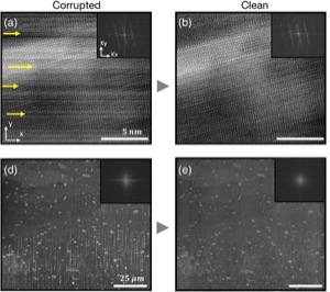
Highly-directional image artifacts such as ion mill curtaining, mechanical scratches, or image striping from beam instability degrade the interpretability of micrographs. These unwanted, aperiodic features extend the image along a primary direction and occupy a small wedge of information in Fourier space. Deleting this wedge of data replaces stripes, scratches, or curtaining, with more complex streaking and blurring artifacts—known within the tomography community as "missing wedge" artifacts. Here, we overcome this problem by recovering the missing region using total variation minimization, which leverages image sparsity-based reconstruction techniques—colloquially referred to as compressed sensing (CS)—to reliably restore images corrupted by stripe-like features. Our approach removes beam instability, ion mill curtaining, mechanical scratches, or any stripe features and remains robust at low signal-to-noise. The success of this approach is achieved by exploiting CS's inability to recover directional structures that are highly localized and missing in Fourier Space.
@article{osti_1572085,
  title = {Removing Stripes, Scratches, and Curtaining with Nonrecoverable Compressed Sensing},
  author = {Schwartz, Jonathan and Jiang, Yi and Wang, Yongjie and Aiello, Anthony and Bhattacharya, Pallab and Yuan, Hui and Mi, Zetian and Bassim, Nabil and Hovden, Robert},
  journal = {Microscopy and Microanalysis},
  volume = {25},
  number = {3},
  pages = {705--710},
  year = {2019},
  doi = {10.1017/S1431927619000254},
}