I earned my Ph.D. in Material Sciences at the University of Michigan, Ann Arbor advised by Prof. Robert Hovden My Ph.D. research primarily focused on material discovery through image processing, data analysis, and 3D reconstruction of nano- and atomic-scale images collected by electron microscopes. Throughout my Ph.D., I tackled inverse problems in electron microscopy by leveraging concepts from signal processing and deep learning. My current work emphasizes the computational aspects of the field, including algorithm development and optimization, large-scale implementation via cluster computing, and scientific software development.
Research
My research focuses on the intersection of deep learning, computer vision, and scientific imaging. I develop supervised learning and interactive human-in-the-loop workflows, often leveraging foundation models for both 2D and 3D data. These methodologies originated in my materials science work and are now being applied to biological imaging—particularly in cryo-electron microscopy (Cryo-EM) and cryo-electron tomography (Cryo-ET) at the Biohub.
Inverse problem optimization for computational imaging.
During my PhD, I developed computational methods for solving ill-posed inverse problems in electron microscopy, where the goal is to reconstruct high-fidelity 3D structures from incomplete and noisy measurements. My work focused on designing optimization algorithms that leverage complementary information across multiple imaging modalities and measurement geometries.
Deep learning for 3D annotation.
I develop data-efficient learning strategies for large volumetric cryo-ET datasets, where dense 3D supervision is impractical. My work reframes annotation as sparse interaction: experts label a small number of 2D slices, and foundation models propagate those signals consistently throughout 3D volumes. In parallel, I design supervised 3D convolutional networks for protein localization and object detection, addressing the complementary problem of identifying discrete molecular coordinates in noisy cellular environments.
Interactive scientific software.
I translate machine learning and reconstruction methods into scalable, reproducible systems for structural biology. I build end-to-end computational infrastructure that runs reliably on HPC clusters while remaining accessible through interactive, web-based dashboards. By integrating large-scale computation with real-time visualization and workflow monitoring, I shorten the feedback loop between model development, evaluation, and biological discovery..
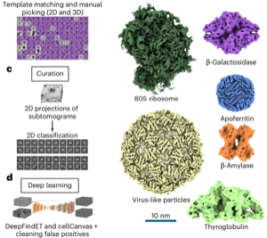
Cryo-electron tomography (cryo-ET) is a powerful technique for imaging molecular complexes in their native cellular environments. However, identifying the vast majority of molecular species in cellular tomograms remains prohibitively difficult. Machine learning (ML) methods provide an opportunity to automate the annotation process, but algorithm development has been hindered by the lack of large, standardized datasets. Here we present an experimental phantom dataset with comprehensive ground-truth annotations for six molecular species to spur new algorithm development and benchmark existing tools. This annotated dataset is available on the CryoET Data Portal with infrastructure to streamline access for methods developers across fields.
@article{peck2025phantom,
  title = {A Realistic Phantom Dataset for Benchmarking Cryo-ET Data Annotation},
  author = {Peck, Ariana and Yu, Yue and Schwartz, Jonathan and Cheng, Anchi and Ermel, Utz Heinrich and Hutchings, Joshua and Kandel, Saugat and Kimanius, Dari and Montabana, Elizabeth A. and Serwas, Daniel and Siems, Hannah and Wang, Feng and Zhao, Zhuowen and Zheng, Shawn and Haury, Matthias and Agard, David A. and Potter, Clinton S. and Carragher, Bridget and Harrington, Kyle and Paraan, Mohammadreza},
  journal = {Nature Methods},
  volume = {22},
  number = {9},
  pages = {1819--1823},
  year = {2025},
  doi = {10.1038/s41592-025-02800-5},
}
Inverse Problems
Imaging 3D Chemistry at 1 nm Resolution with Fused Multi-Modal Electron Tomography
Jonathan Schwartz, Zichao Wendy Di, Yi Jiang, and
14 more authors
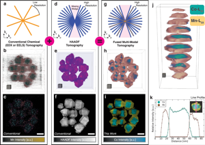
Measuring the three-dimensional (3D) distribution of chemistry in nanoscale matter is a longstanding challenge for metrological science. The inelastic scattering events required for 3D chemical imaging are too rare, requiring high beam exposure that destroys the specimen before an experiment is completed. Even larger doses are required to achieve high resolution. Thus, chemical mapping in 3D has been unachievable except at lower resolution with the most radiation-hard materials. Here, high-resolution 3D chemical imaging is achieved near or below one-nanometer resolution in an Au-Fe3O4 metamaterial within an organic ligand matrix, Co3O4-Mn3O4 core-shell nanocrystals, and ZnS-Cu0.64S0.36 nanomaterial using fused multi-modal electron tomography. Multi-modal data fusion enables high-resolution chemical tomography often with 99% less dose by linking information encoded within both elastic (HAADF) and inelastic (EDX/EELS) signals. We thus demonstrate that sub-nanometer 3D resolution of chemistry is measurable for a broad class of geometrically and compositionally complex materials.
@article{yang2024imaging3d,
  title = {Imaging 3D Chemistry at 1 nm Resolution with Fused Multi-Modal Electron Tomography},
  author = {Schwartz, Jonathan and Di, Zichao Wendy and Jiang, Yi and Manassa, Jason and Pietryga, Jacob and Qian, Yiwen and Cho, Min Gee and Rowell, Jonathan L. and Zheng, Huihuo and Robinson, Richard D. and Gu, Junsi and Kirilin, Alexey and Rozeveld, Steve and Ercius, Peter and Fessler, Jeffrey A. and Scott, Mary and Hovden, Robert},
  journal = {Nature Communications},
  volume = {15},
  pages = {47558},
  year = {2024},
  doi = {10.1038/s41467-024-47558-0},
}
Sci. Software
Real-time 3D Analysis During Electron Tomography Using tomviz
Jonathan Schwartz, Chris Harris, Jacob Pietryga, and
14 more authors
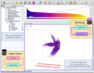
The demand for high-throughput electron tomography is rapidly increasing in biological and material sciences. However, this 3D imaging technique is computationally bottlenecked by alignment and reconstruction which runs from hours to days. We demonstrate real-time tomography with dynamic 3D tomographic visualization to enable rapid interpretation of specimen structure immediately as data is collected on an electron microscope. Using geometrically complex chiral nanoparticles, we show volumetric interpretation can begin in less than 10 minutes and a high-quality tomogram is available within 30 minutes. Real-time tomography is integrated into tomviz, an open-source and cross-platform 3D data analysis tool that contains intuitive graphical user interfaces (GUI), to enable any scientist to characterize biological and material structure in 3D.
@article{schwartz2022real,
  title = {Real-time 3D Analysis During Electron Tomography Using tomviz},
  author = {Schwartz, Jonathan and Harris, Chris and Pietryga, Jacob and Zheng, Huihuo and Kumar, Prashant and Visheratina, Anastasiia and Kotov, Nicholas A. and Major, Brianna and Avery, Patrick and Ercius, Peter and Ayachit, Utkarsh and Geveci, Berk and Muller, David A. and Genova, Alessandro and Jiang, Yi and Hanwell, Marcus and Hovden, Robert},
  journal = {Nature Communications},
  volume = {13},
  number = {1},
  pages = {4458},
  year = {2022},
  doi = {10.1038/s41467-022-32046-0},
}
Inverse Problems
Imaging Atomic-Scale Chemistry from Fused Multi-Modal Electron Microscopy
Jonathan Schwartz, Zichao Wendy Di, Yi Jiang, and
9 more authors
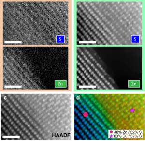
Efforts to map atomic-scale chemistry at low doses with minimal noise using electron microscopes are fundamentally limited by inelastic interactions. Here, fused multi-modal electron microscopy offers high signal-to-noise ratio (SNR) recovery of material chemistry at nano- and atomic-resolution by coupling correlated information encoded within both elastic scattering (high-angle annular dark-field (HAADF)) and inelastic spectroscopic signals (electron energy loss (EELS) or energy-dispersive x-ray (EDX)). By linking these simultaneously acquired signals, or modalities, the chemical distribution within nanomaterials can be imaged at significantly lower doses with existing detector hardware. In many cases, the dose requirements can be reduced by over one order of magnitude. This high SNR recovery of chemistry is tested against simulated and experimental atomic resolution data of heterogeneous nanomaterials.
@article{mi2022imaging,
  title = {Imaging Atomic-Scale Chemistry from Fused Multi-Modal Electron Microscopy},
  author = {Schwartz, Jonathan and Di, Zichao Wendy and Jiang, Yi and Fielitz, Alyssa J. and Ha, Don-Hyung and Perera, Sanjaya D. and El Baggari, Ismail and Robinson, Richard D. and Fessler, Jeffrey A. and Ophus, Colin and Rozeveld, Steve and Hovden, Robert},
  journal = {npj Computational Materials},
  volume = {8},
  pages = {16},
  year = {2022},
  doi = {10.1038/s41524-021-00692-5},
}
Inverse Problems
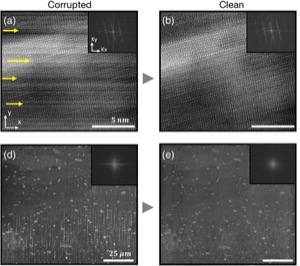
Removing Stripes, Scratches, and Curtaining with Nonrecoverable Compressed Sensing
Jonathan Schwartz, Yi Jiang, Yongjie Wang, and
6 more authors
Highly-directional image artifacts such as ion mill curtaining, mechanical scratches, or image striping from beam instability degrade the interpretability of micrographs. These unwanted, aperiodic features extend the image along a primary direction and occupy a small wedge of information in Fourier space. Deleting this wedge of data replaces stripes, scratches, or curtaining, with more complex streaking and blurring artifacts—known within the tomography community as "missing wedge" artifacts. Here, we overcome this problem by recovering the missing region using total variation minimization, which leverages image sparsity-based reconstruction techniques—colloquially referred to as compressed sensing (CS)—to reliably restore images corrupted by stripe-like features. Our approach removes beam instability, ion mill curtaining, mechanical scratches, or any stripe features and remains robust at low signal-to-noise. The success of this approach is achieved by exploiting CS's inability to recover directional structures that are highly localized and missing in Fourier Space.
@article{osti_1572085,
  title = {Removing Stripes, Scratches, and Curtaining with Nonrecoverable Compressed Sensing},
  author = {Schwartz, Jonathan and Jiang, Yi and Wang, Yongjie and Aiello, Anthony and Bhattacharya, Pallab and Yuan, Hui and Mi, Zetian and Bassim, Nabil and Hovden, Robert},
  journal = {Microscopy and Microanalysis},
  volume = {25},
  number = {3},
  pages = {705--710},
  year = {2019},
  doi = {10.1017/S1431927619000254},
}